
image captured with TelScope Telehealth System
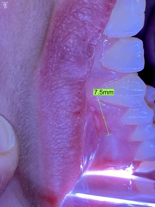
image captured with TelScope Telehealth System
PROBLEM
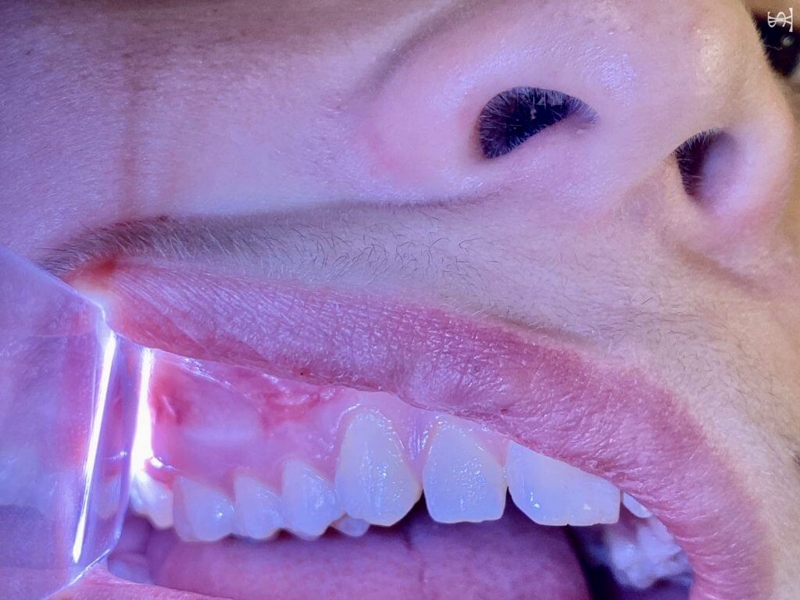
A 12-year-old male complains of pain for three days. His mother is concerned that he has an “infection in his gums.” A clinical exam performed in the dental office was documented using a TelScope. The first image reveals two oral lesions above the teeth and gum tissue in the upper right quadrant, located in what is called unattached mucosa. The appearance of a white center surrounded by a red halo is similar to an aphthous ulcer (canker sore), but the shape of the larger lesion is not typical of a mouth ulcer, which is usually more ovoid.
Careful extra-oral examination reveals an area of contusion (bruising) of the upper lip near the sores. Directly behind this discolored area is the irregular, linear oral lesion measuring approximately 7.5mm. A smaller lesion is located more distal.
Questioning the patient provided the answers to the mystery. Three to four days earlier, the patient was chewing on a small straw and he was bumped in the mouth by another child.
DIAGNOSIS
The typical appearance is a whitish-yellow central area surrounded by an inflammatory red (erythematous) halo, often on a rolled border. They are painful, and “sting” when exposed to high acid products such as ketchup, tomato based sauces, and citrous products. They typically heal in 10-14 days. Ibuprofen can help with pain if needed, and an over the counter oral paste can be used to cover the ulceration to improve comfort if desired. Your dentist can also prescribe an oral paste with a topical steroid.
COMMENTARY

About the Author: Dr. Richard Simpson, DMD
INFORMATION & CONTENT DISCLAIMER
This content is for information only. This content is not for advice, diagnosis, or
guarantee of outcome for patients. No patients should use the information,
resources, or tools contained within to self-diagnose or self-treat any health-related
conditions.