Facial Swelling Caused by Dental Infection – by Dr. Richard Simpson, DMD

facial swelling

image captured with TelScope Telehealth System

PROBLEM

A 5-year-old male was seen in a medical emergi-care center with a chief complaint from the parent of pain and “his face is swollen”. He was also febrile (showing symptoms of a fever). The patient was diagnosed with a rapidly developing facial cellulitis (infection spreading into the soft tissues of the face), of probable dental origin. The child was given a shot of Rocephin, and was prescribed an oral antibiotic and an over-the-counter pain medication. The patient was then given a dental referral for further evaluation and treatment.
Note: Untreated facial cellulitis from a dental infection can lead to multiple systemic health complications, blindness, difficulty breathing, and in rare cases, death.

DIAGNOSIS

Upon dental exam, the facial swelling had resolved in response to the antibiotics. This intraoral image reveals the swelling was now localized and associated with an upper left second primary (“baby”) molar that had a large cavity (dental caries) present. This presentation and diagnosis is considered urgent, and treatment should be rendered as soon as possible. The tooth will require extraction (removal). A discussion of the recommendation for a space maintainer should take place.

INTER-PROFESSIONAL COLLABORATION FOR IDEAL PATIENT CARE

This case is an excellent example of how healthcare providers, equipped with a TelScope in their facility, are able to triage a patient and obtain a teledentistry consult for an initial diagnosis and treatment plan, followed by a referral for appropriate treatment. This inter-professional collaboration through telehealth capabilities leads to effective and efficient health care, and can reduce appointment numbers, time, and exposure to others during these times of COVID-19.

About the Author: Dr. Richard Simpson, DMD

Dr. Richard is a board certified pediatric dentist in private practice.
His achievements include:
• Diplomate in the American Board of Pediatric Dentistry
• Fellow in the American College of Dentists
• Fellow in the International College of Dentists
• Fellow in the American Academy of Pediatric Dentistry
• Advisory Board Member of The TeleDentists
• Advisory Board Member of Holland Healthcare
Dr. Richard’s experience and active interests include telehealth, child advocacy, health disparities, policy, and advancing improved medical-dental access to care. Dr. Richard is also a veteran with 15 years of military service.
————
INFORMATION & CONTENT DISCLAIMER

This content is for information only. This content is not for advice, diagnosis, or
guarantee of outcome for patients. No patients should use the information,
resources, or tools contained within to self-diagnose or self-treat any health-related
conditions. 

Sign up for more content like this from Holland Healthcare:

Subscribe

* indicates required

Profession / Field

Which upcoming device(s) are you interested in?

Chipped Tooth as a Sign of a Cavity – by Dr. Richard Simpson, DMD

Chipped tooth

image captured with TelScope Telehealth System

Chipped tooth

image captured with TelScope Telehealth System

PROBLEM

A 6-year-old male reported to the dental office with his mother, who stated she noticed “a chipped tooth”. There was no current pain reported. However, after questioning, it was confirmed that the patient had experienced oral pain for “a few days” several weeks earlier, but it went away.

DIAGNOSIS

Clinical exam and an x-ray confirmed the patient had a large cavity (dental caries) in the lower right first primary molar. The second photograph, marked using the TelScope App, shows a fistula that developed as a result of a previous tooth abscess. This allowed the infection to eventually drain, which resolved the patient’s original pain. The nerve of the tooth is no longer vital (alive). This is a non-urgent diagnosis, but treatment is indicated. The treatment consists of extraction of the primary tooth, as well as a recommendation for a space maintainer to be placed on the second molar to hold eruption space for the replacement tooth, which usually emerges at age ten to eleven. Failure to remove the tooth could lead to multiple complications to include further fracturing of the tooth, space loss, shifting of teeth, recurrence of the infection, or damage to the underlying permanent tooth.

INTERESTING FACT

Tooth decay can be categorized by the type of tooth surface and the location on the tooth in which it develops. This cavity developed between the teeth and was originally much smaller. An x-ray taken at a routine dental exam can identify this type of cavity before it is visible in the mouth. Early diagnosis is important, because dental decay in primary teeth (“baby teeth”) typically increases in size four to six times faster than in permanent teeth.

About the Author: Dr. Richard Simpson, DMD

Dr. Richard is a board certified pediatric dentist in private practice.
His achievements include:
• Diplomate in the American Board of Pediatric Dentistry
• Fellow in the American College of Dentists
• Fellow in the International College of Dentists
• Fellow in the American Academy of Pediatric Dentistry
• Advisory Board Member of The TeleDentists
• Advisory Board Member of Holland Healthcare
Dr. Richard’s experience and active interests include telehealth, child advocacy, health disparities, policy, and advancing improved medical-dental access to care. Dr. Richard is also a veteran with 15 years of military service.
————
INFORMATION & CONTENT DISCLAIMER

This content is for information only. This content is not for advice, diagnosis, or
guarantee of outcome for patients. No patients should use the information,
resources, or tools contained within to self-diagnose or self-treat any health-related
conditions. 

Sign up for more content like this from Holland Healthcare:

Subscribe

* indicates required

Profession / Field

Which upcoming device(s) are you interested in?

Identifying an Oral Lesion | Dr. Richard Simpson, DMD

Oral lesion

image captured with TelScope Telehealth System

Oral lesion

image captured with TelScope Telehealth System

PROBLEM

A 6-year-old female patient was seen for a routine Well Child exam by her pediatrician. The doctor noted a “swelling” on her lower lip, and the mother reported that it had been present for 4 weeks. The physician recommended a teledentistry evaluation with a dentist and referred her.

DIAGNOSIS

The “swelling” in question appeared as a raised, 4mm x 3mm soft tissue lesion with a broad base and normal color. It is located in the midline of the lower lip inside of the wet-dry line. The lesion did not change in size since it first appeared, and was not painful.
The diagnosis is a mucocele. A mucocele is a harmless fluid-filled retention “cyst” that most commonly results from the rupture of a minor salivary gland. Hundreds of salivary glands are located under the surface of the lip and other areas of the oral mucosa to maintain moisture. If a mucocele does not disappear after 4-6 weeks, it should be removed by a dentist or an oral surgeon, as it can interfere with normal function and often lead to the development of chronic biting or sucking habits and scar tissue, especially in children.
This is an excellent example of how an oral lesion or area of concern could be pre-screened by a healthcare provider, photographed using the TelScope Telehealth System, and transmitted to a dentist for a teledentistry consult prior to scheduling an appointment.

About the Author: Dr. Richard Simpson, DMD

Dr. Richard is a board certified pediatric dentist in private practice.
His achievements include:
• Diplomate in the American Board of Pediatric Dentistry
• Fellow in the American College of Dentists
• Fellow in the International College of Dentists
• Fellow in the American Academy of Pediatric Dentistry
• Advisory Board Member of The TeleDentists
• Advisory Board Member of Holland Healthcare
Dr. Richard’s experience and active interests include telehealth, child advocacy, health disparities, policy, and advancing improved medical-dental access to care. Dr. Richard is also a veteran with 15 years of military service.
————
INFORMATION & CONTENT DISCLAIMER

This content is for information only. This content is not for advice, diagnosis, or
guarantee of outcome for patients. No patients should use the information,
resources, or tools contained within to self-diagnose or self-treat any health-related
conditions. 

Sign up for more content like this from Holland Healthcare:

Subscribe

* indicates required

Profession / Field

Which upcoming device(s) are you interested in?


Eruption of a Permanent Tooth – by Dr. Richard Simpson, DMD

permanent tooth eruption

image captured with TelScope Telehealth System

Permanent tooth eruption

image captured with TelScope Telehealth System

PROBLEM

10-year-old female patient says, “I feel something in the top of my mouth”. No pain reported.

DIAGNOSIS

A clinical exam, documented here with a TelScope image, confirms an over retained primary (baby) tooth with a stainless steel crown present.

It’s replacement permanent tooth is emerging (erupting) in the palate. The primary tooth was not mobile because a portion of the roots failed to resorb. Although this is not an urgent situation, the removal of the primary tooth by a dentist is indicated to allow the permanent tooth to fully erupt. The permanent tooth will likely drift back toward a more ideal position if the baby tooth is removed soon.

INTERESTING FACT

This is not an uncommon problem, and is an excellent example of a situation that can be pre-screened by a dentist through a teledentistry consultation with a clear photograph such as this that can easily be taken with a TelScope Telehealth System.

About the Author: Dr. Richard Simpson, DMD

Dr. Richard is a board certified pediatric dentist in private practice.
His achievements include:
• Diplomate in the American Board of Pediatric Dentistry
• Fellow in the American College of Dentists
• Fellow in the International College of Dentists
• Fellow in the American Academy of Pediatric Dentistry
• Advisory Board Member of The TeleDentists
• Advisory Board Member of Holland Healthcare
Dr. Richard’s experience and active interests include telehealth, child advocacy, health disparities, policy, and advancing improved medical-dental access to care. Dr. Richard is also a veteran with 15 years of military service.
————
INFORMATION & CONTENT DISCLAIMER

This content is for information only. This content is not for advice, diagnosis, or guarantee of outcome for patients. No patients should use the information, resources, or tools contained within to self-diagnose or self-treat any health-related conditions. 

Sign up for more content like this from Holland Healthcare:

Subscribe

* indicates required

Profession / Field

Which upcoming device(s) are you interested in?


Dental Growing Pains – by Dr. Richard Simpson, DMD

dental growing pains

PROBLEM

An 11-year-old female patient is complaining of occasional pain in the lower right quadrant of her mouth in the evenings and at night for two weeks now.  The patient’s mother is concerned about possible “swelling”. This teledentistry photo taken with TelScope Telehealth System shows there is a permanent second molar emerging (“erupting”) in a normal position.

DIAGNOSIS

The tissue over the back of the tooth will recede with time as the tooth erupts into the mouth further. There is no inflammation or infection, and no treatment is indicated. The patient’s mother was instructed that the patient should brush the emerging tooth as normal. She could give her child an over the counter pain medication like acetaminophen or ibuprofen if needed.

INTERESTING FACT

Children grow, and teeth move, in the evenings and nighttime, as their growth hormone increases during this time. It is not uncommon to occasionally have discomfort from tooth movement that may come and go with no other symptoms – dental “growing pains”.

However, other more significant oral health issues can also cause night time pain. A photograph and a teledentistry consult with a dentist can help determine if an in-office examination is indicated.

About the Author: Dr. Richard Simpson, DMD

Dr. Richard is a board certified pediatric dentist in private practice.
His achievements include:
• Diplomate in the American Board of Pediatric Dentistry
• Fellow in the American College of Dentists
• Fellow in the International College of Dentists
• Fellow in the American Academy of Pediatric Dentistry
• Advisory Board Member of The TeleDentists
• Advisory Board Member of Holland Healthcare
Dr. Richard’s experience and active interests include telehealth, child advocacy, health disparities, policy, and advancing improved medical-dental access to care. Dr. Richard is also a veteran with 15 years of military service.
————
INFORMATION & CONTENT DISCLAIMER

This content is for information only. This content is not for advice, diagnosis, or
guarantee of outcome for patients. No patients should use the information,
resources, or tools contained within to self-diagnose or self-treat any health-related
conditions. 

Sign up for more content like this from Holland Healthcare:

Subscribe

* indicates required

Profession / Field

Which upcoming device(s) are you interested in?


How to Capture Amazing Oral Telehealth & Teledentistry Images with TelScope Telehealth System

How to Get an Amazing Oral Telehealth or Teledentistry Image with TelScope Telehealth System

To get an image with the TelScope Telehealth System for telehealth or teledentistry, first, examine the patient with the TelScope handle and single-use depressor before attaching it to the smart device.

Be sure to depress the tongue to illuminate inside the mouth.

For teledentistry and to get a good view of the molars and the teeth, retract the cheek and slide the TelScope sideways between the gum and the cheek.

If you see something of interest, proceed to attach TelScope to your smart device. Open the TelScope app to capture an intraoral telehealth or teledentistry image.

  • The single-use depressor should be attached to the TelScope handle before attaching the TelScope handle to your smart device

Now capture an amazing telehealth or teledentistry image.

The TelScope app is automatically set with optimal lighting, focus, and zoom settings for superior quality intraoral imagery.

The TelScope app camera is automatically zoomed in to focus on the inside of the mouth. We do not recommend zooming in or out except for capturing specific images.

Place the TelScope blade inside your patient’s mouth and depress the tongue to illuminate inside the mouth. 

A good indication of a well-lit mouth is when the uvula is illuminated. 

For teledentistry images, turn your phone sideways, retract the cheek, and slide the depressor sideways between the cheek and the gum. This technique will allow you to capture images of the molars and the teeth.

We recommend practicing with TelScope on yourself in the mirror to get the hang of how to capture amazing images.

TelScope App Best Practices

• Make sure you are using a HIPAA compliant smart device.

• Images should be uploaded to your patient file or sent to a peer for review and then deleted from the app directly after your patient’s appointment.

• Capture multiple images during the patient exam, then choose the best images from the app to upload to the patient file. Delete images from the app after upload.

• For measurement indications, your TelScope metal clip must be accurately placed and depressor visible in the photo.

Watch a TelScope Telehealth System Demonstration with Inventor Jennifer Holland:

Still need help? 

Reach out to Mike at mvogel@hollandhealthcareinc.com to schedule a free TelScope live tutorial!

Introducing TelScope Oral Telehealth System

Introducing TelScope Oral Telehealth System

Introducing the telehealth version of Throat Scope: TelScope Oral Telehealth System.

Connect intraoral concerns to a doctor or specialist quickly without leaving home, or have clients connect directly to you.

TelScope is a product and app combination. The TelScope handle is like the upgraded Throat Scope – it’s antimicrobial, rechargeable, and connects to any smart device.

When you connect TelScope to your smart device, open the TelScope app (available on iOS and Android) to snap an intraoral picture. Then you can measure, circle and comment on any areas of concern. Finally, send the image instantly to a doctor, dentist or specialist securely within the app.


TelScope Oral Telehealth System TelScope Oral Telehealth System

You can learn more about TelScope on our parent company website here.

TelScope is ready for preorder – for an amazing discount – via Indiegogo. Preorder here.


What People Are Saying About Throat Scope

Dentist Oral Hygienist using Throat Scope Illuminating Tongue Depressor with Child

What are people saying about Throat Scope? Pediatricians, general practitioners, primary care physicians, dentists, speech-language pathologists and more across 155 countries are using Throat Scope. Read reviews from professionals across all scopes of practice.

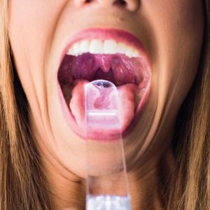
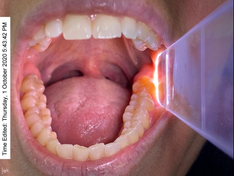
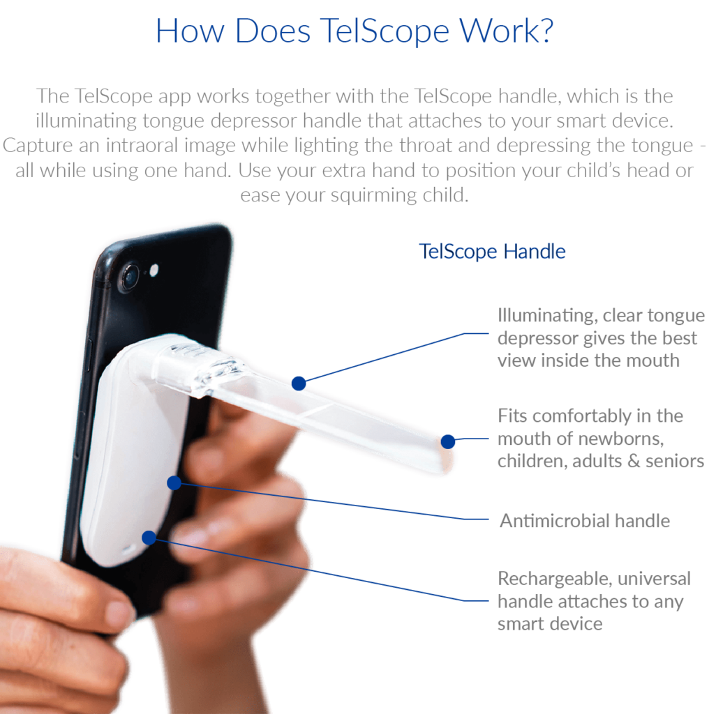
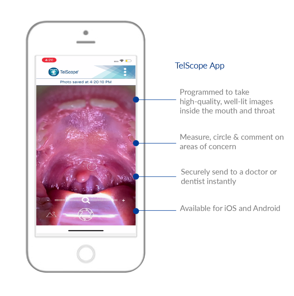
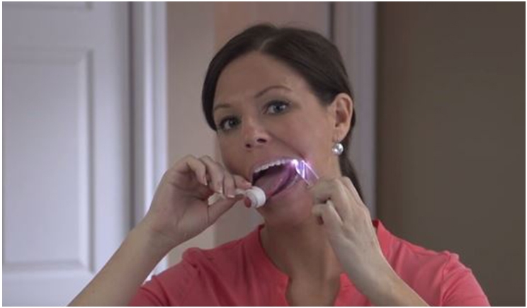
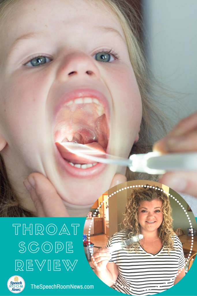
Throat Scope makes experiences for yourself and your patient faster, easier, and most importantly – more accurate. Throat Scope frees up an extra hand instead of the old flashlight-depressor combo by placing the light source directly inside the mouth.

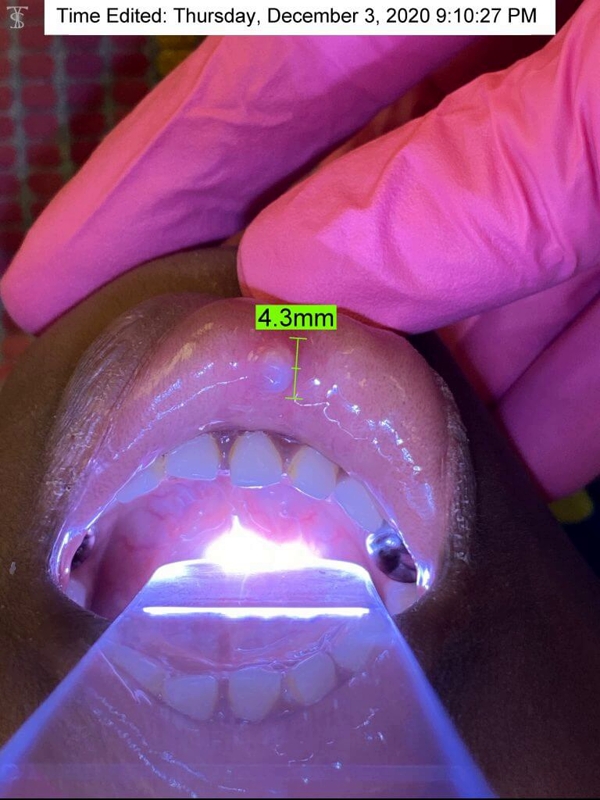
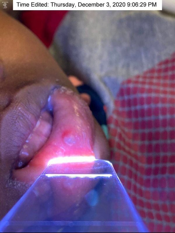
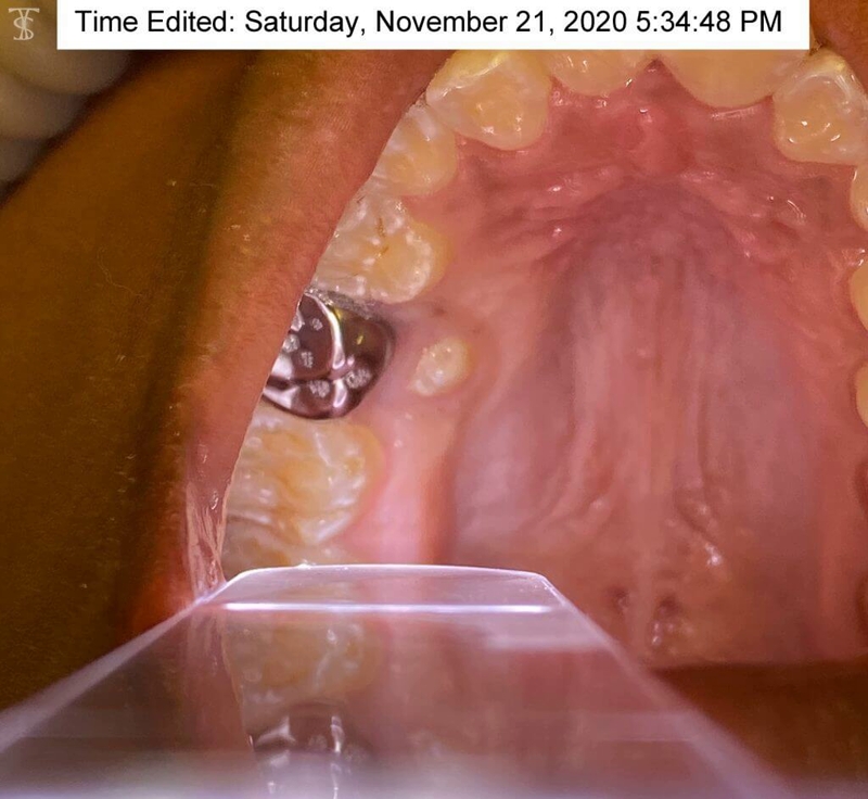
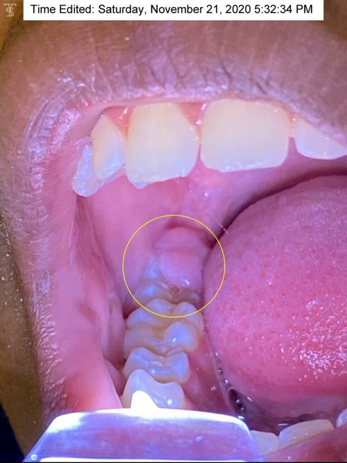
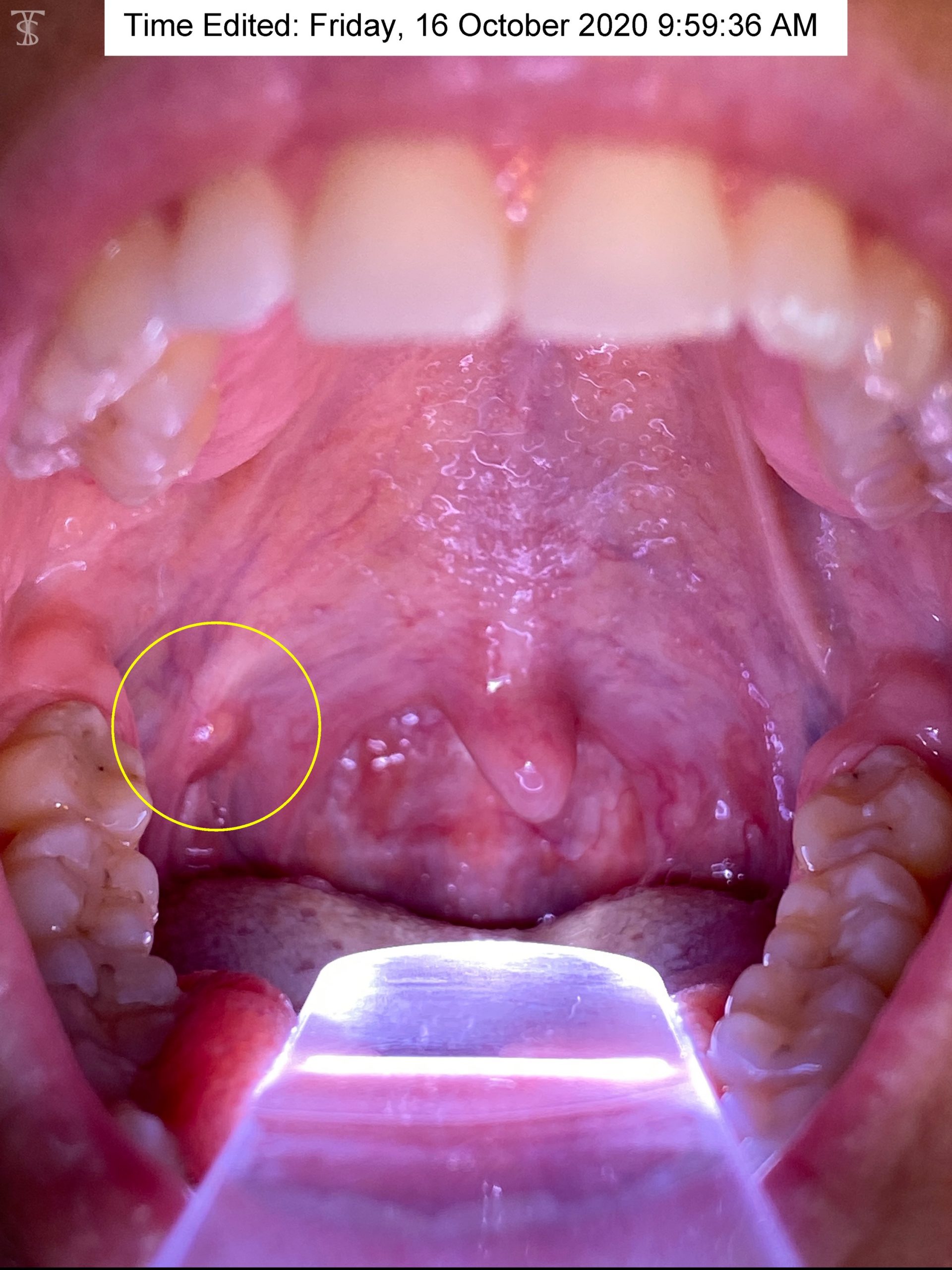
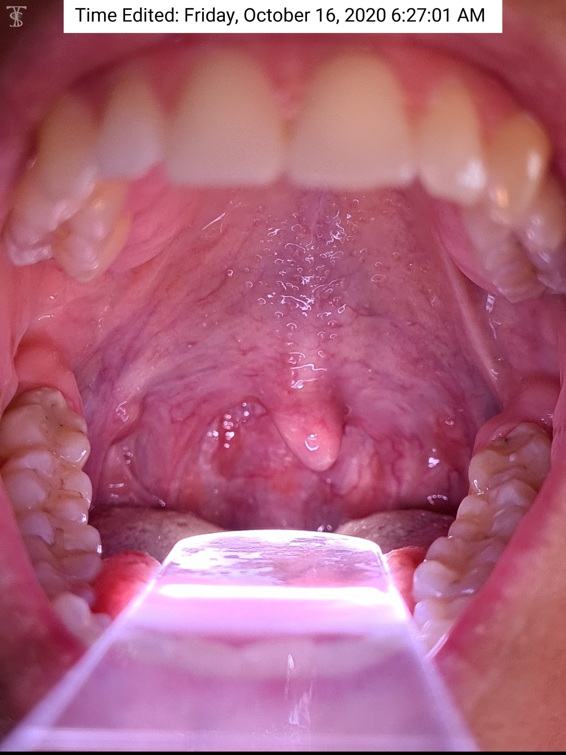
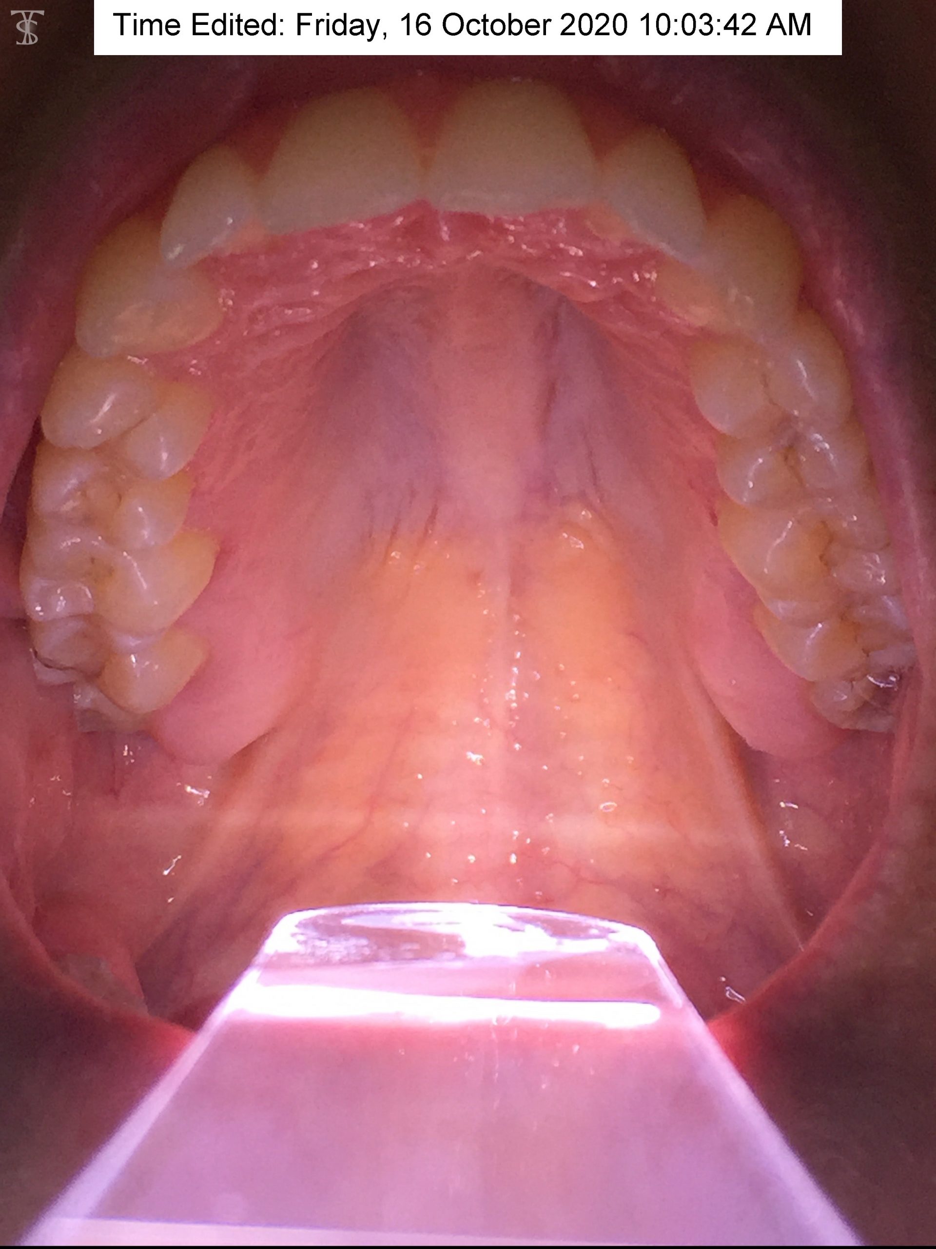
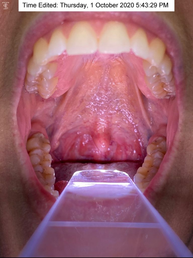
Throat Scope is also used as a speech and articulation tool, an examination tool, and an oral cancer screening tool.

Here’s what people are saying about Throat Scope:

“This is an absolute essential for every dental and medical practitioner to provide the best possible oral assessment and ability to discover oral cancer in its earliest stages.”
Jo-Anne Jones – Dental Hygienist & Oral Cancer Motivational Speaker

:This is a product that I have been using like crazy with a few of my /k/ and /g/ kids, actually all of my kids. It also looks like a light saber which makes every kid want to do it. Finally a therapy tool we can afford!”
Jenna – Speech Language Pathologist – Speech Room News

“I’m a school nurse and with all the strep and flu going around, I am going to fly through my box of 100!
Throat Scope is life changing for me! It makes checking kids a breeze. I am awestruck! I’m a “one man band” it’s singing praises to professionals and family and friends. Brilliant!”
Paula – School Nurse

“We are loving using our new Throat Scopes in the clinic. What a difference it makes to be able to see inside our ‘littlies’ mouths quickly and easily. What a great idea! Also very useful for identifying air escaping down the nose during speech for our Cleft Palate clients as it so happens!”
Kim – Speech Language Pathologist

“Throat Scope is high-tech compared to the organic incumbent.”
ZD Net – Worldwide News Media

“Throat Scope provides medical professionals with one free hand, a light source located inside the mouth for a fast, accurate and pleasant oral examination experience.”
The Daily Floss Worldwide News Media

One word: Genius.
SLP Toolkit – Speech Language Pathologist

“It lights up the whole mouth! You can see a lot more than redness at the back of the mouth #brilliant
Eva Grayzel – Motivational Speaker for Oral Cancer

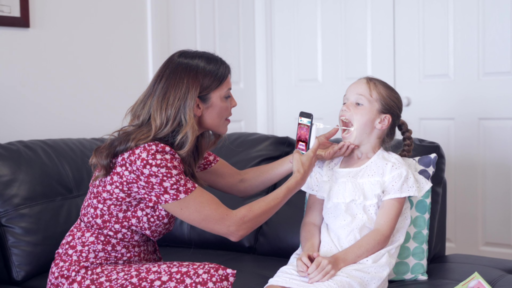
“Parents and school nurses who have students with special needs who have trouble getting their kids open their mouths long enough to see what going in the back of their throat. At least with Throat Scope, you get a clear look even if it is quick!”
Carla Butorac – Speech Language Therapist

“This award comes with a lot of “firsts”. While only a couple of Australian companies have been honoured with an Edison Award since their inception in 1987, Holland Healthcare is the first Australian female led company to be recognised. It is also the first Australian medical device to win.”
Hunter Headline Newcastle Newspaper

Cannot believe my daughter did not gag. She was so excited to have Throat Scope that she didn’t mind the usual discomfort that goes along with an oral examination!”
Katy – Speech Pathology Mama

“No more juggling with a torch and a wooden craft stick to look into a kid’s gob. It’s a blessing for clinicians and parents. Who knew oral cavity examinations could be so much fun?”
The Australian Newspaper

“The Throat Scope is great. More kid friendly than a traditional tongue depressor and pen-torch, for assessing children’s mouth and throat.”
Kathryn Harber – Speech Language Pathologist

“This product is well made and arrived really quickly. I love the product and it’s been very useful already.”
Melody Humphries- Speech Language Pathologist

“It works, reduces the materials I need, and engages kids. It’s a win.”
Jenna – Speech Language Pathologist – Speech Room News

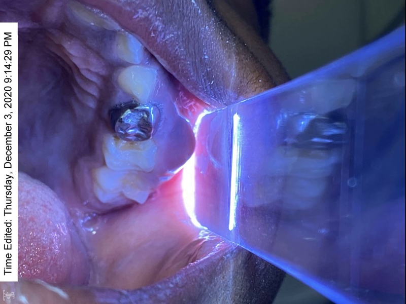
“Having another ‘tool’ to help illuminate challenging areas of the mouth including the tongue, floor of the mouth, palate and throat makes earlier discovery more feasible. Great adjunct for screening examinations and oral self exams.”
Jo-Anne Jones – Motivational Speaker on Oral Cancer

“This is an SLP’s dream!”
Amanda Schaumburg – Speech Language Pathologist – Panda Speech

“My kids love it, it has a light saber element to it.”
Nacole – Registered Nurse – NurseNacole
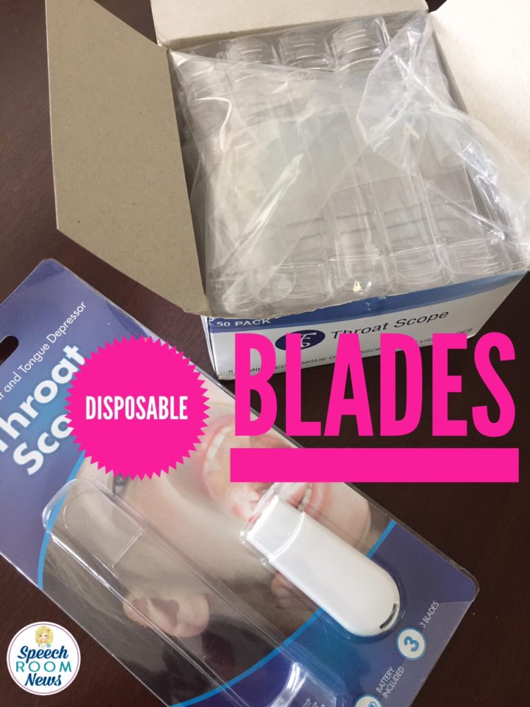
I have 3 of these scopes, one in each exam room. I purchased 150 count of the blades. I use them not only in assessment but in treatment with patients. I place one blade with each patient for sensory targets, or useful in resistive exercises and in demonstrations of therapeutic exercises-side by side.
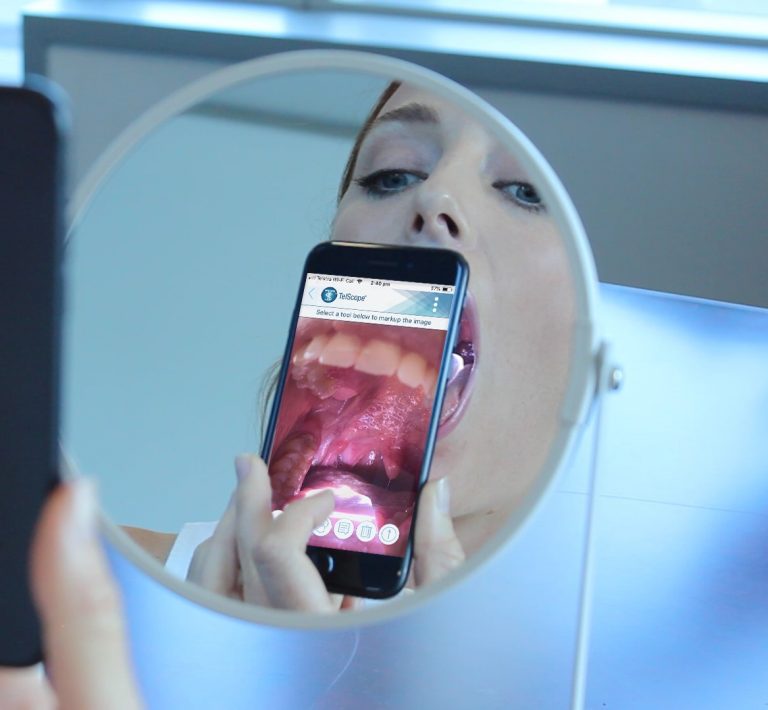
I also use these with my current cancer patients undergoing radiation treatment . I am able to take photographic images for communication with the radio-oncologist and or ENT to quickly manage mucositis; thrush and lesion development.”
Tamatha Rutherford – Speech Language Pathologist

“This is a great product especially for patients with structural abnormalities, oral apraxia or macroglossia which can make visualizing the oral cavity a real challenge! Much more effective than using tongue depressor in one hand and pen light in the other!
Anne Brockman – Speech Language Pathologist

“Throat Scope is a game changer for OME and oral exams”
Graham Speech Therapy – Speech Language Pathologist

Best Intraoral Exam Tool Ever.
Dr Keith Grimes – UK Doctor

“My area of specialization in dermatology is blistering diseases. Patients may have erosions and blisters inside the mouth as well as other mucous membranes and of course, on the skin. During these patient reviews, I have found it time saving and patient friendly to wear the lanyard which attaches to a device called a ‘throat scope’ which contains a battery-powered light. There is a disposable see-through plastic tongue depressor which is individually wrapped that I attach to this in order to easily view inside the mouth and take photos with my free hand. It is also useful for examining the mouth for skin cancers and infections.”
Dedee F. Murrell, Professor of Dermatology, University of Sydney, Australia

“I could be a salesperson for Throat Scope. I love this tool and use it daily as a clinician . As a clinical teacher, I’m constantly introducing it to the next generation of SLPs. Not only is it functional, but it is also fun. A” must have ” when working with pediatrics.”

“Received my delivery today (arrived within days of placing my order). Had a little play around and am very impressed with the quality of manufacture and ease of use. What a valuable tool to have at home and very affordable. I hope to see my GP using this next time I have to visit.”

“Just received this, and with frequent mouth ulcers I expect it will help. BUT, I ordered a 2nd set for my neighbor who has 15 grandkids, the oldest being 16 yrs old, and the youngest being 2 yrs, 1 yr, and 3 days old. Giving it to her since most of the kids are in and out of her house every single day. Thanks for a simple but genius product!

“Great product! So useful for dental assessments particularly when assessing clients outside the dental clinic. (ie. in ages care facilities) and apprehensive children.”

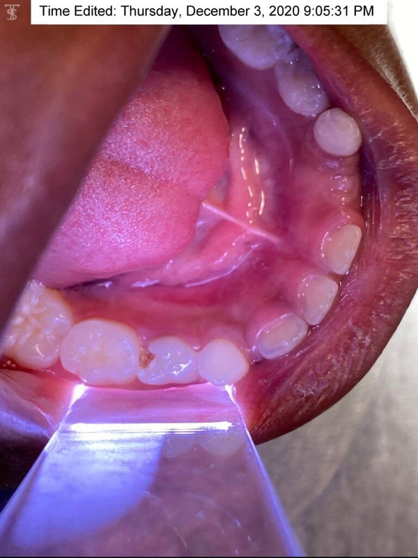
“I love this product – I love this product, all the oral cavity can be seen and even deep into the pharyngeal walls. The light is bright enough and the scope is clear to allow a better view. The throat scope is easy to use, once you connect the clear tongue depressor to the white attachment the light turns on and it’s ready to use. Clear disposable plastics are available and can be placed on the clear tongue depressor for each person.”

“A must have for every SLP! I am a speech-language pathologist and Throat Scope is so helpful for my job! This makes completing oral mech exams so much easier!”

“Came as pictured, words great, very fast. This came exactly as pictured and very quickly! It was super helpful checking my kids after tonsillectomies for healing process and at work for oral mech exams.”

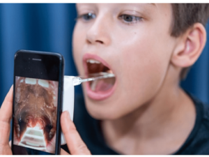
“Good for moms to check their kids throats.”

“These work great. Take it from the Doc, these are worth the cost.”

“Effective to detect problems in the throat. These are very useful when my children get sick.”

“FANTASTIC! This a great product and it performs exactly as advertised. It makes it real easy to see in the persons mouth. I highly recommend it.”

“Great tool! This is an awesome invention. Completing an oral assessment or checking your kids throats or teeth- light and tongue blade in one hand!!!”

“Great product, I even bought one for my doctor!”

“Useful and affordable.”

“I cannot wait to use these in the upcoming school year. This product will make oral examinations much easier than holding a flashlight in one hand and a tongue depressor in the other.”

Activity Tailor – Easier Assessments & Articulation with Throat Scope

Throat Scope is the world’s first all-in-one light and tongue depressor that makes for more accurate and easier assessments and articulation therapy.

Original blog by Kim from Activity Tailor

Oral Mech Exams

Let’s talk assessments and articulation. I need to come clean here. I don’t do a lot of oral mech exams. I’ll do a cursory look at how the tongue is moving and see if the can modify the movement with some verbal cues during assessments, but I’m hesitant to alienate a child I might start seeing for therapy during their assessment by holding their tongue down with a tongue depressor and really getting in there. (And I would never try for a gag reflex! Not with my population who would be much more likely to throw-up on me than give me any neurological insight.)

I do pull out Dum-Dum, flavored tongue depressors and the occasional toothette to help kiddos see or feel where I’m asking them to position their lips/tongues, but… now I’ve got an even better trick up my sleeve.

How can you see what’s actually going on in the mouth during assessments?

The main problem is that it’s so hard to see what’s actually going on in there. I always feel vaguely like I’m spelunking. Which is why I was so keen to try out Throat Scope after I’d seen several photos in my social media feed over the summer and heard all the positive feedback.

What I – and the kiddos – love about Throat Scope

It’s justified. My little ones were so excited about the Throat Scope (which is definitely not what I call it in the speech room–speech light saber is (ironically) much less ominous sounding) and looking in their mouths and looking at all the stuff in there. All of the sudden they were attending to their tongue position, seeing and feeling what I was asking them to do. Makes assessments and articulation exercises much, much easier!

Everyone seemed to find the plastic, even though it’s unflavored, much more palatable than wood tongue depressors.

How does it work?

Here’s how it works. You have a handle with the light inside that you use each time. You buy a box of clear, plastic, disposable depressors that simply slide on to the end (pushing down the toggle and turning the light on). When you’re done, just toss the clear plastic part.

Except most of the kiddos I tried this with begged to keep their “saber” even though they wouldn’t be able to make it illuminate their light. I even had a couple kids bring the saber back to our next session!

To read the original blog by Activity Tailor, head HERE


Never miss a blog!

Subscribe to get updates on new blogs from Throat Scope.


How to Do an Oral-Facial Exam with Throat Scope

Melanie Potock, MA, CCC-SLP and Amy Graham, MA, CCC-SLP show us how to do an oral-facial exam with Throat Scope.


Never miss a blog!

Subscribe to get updates on new blogs from Throat Scope.

Subscribe

* indicates required

Profession / Field


Head and Neck Cancer Screening

Head and neck cancer screening event

Surgeons and oncologists who work in Head and Neck Cancer Multidisciplinary Teams (MDTs) are qualified to do head and neck cancer screening. The process involves asking about possible symptoms, examining the mouth and the top of the throat with a good light source, and palpating the neck for lumps. Screening clinics which run outside traditional hospital environments are a great way to raise awareness of the signs and symptoms of these cancers, especially among people who may never have heard of the disease. Head and neck cancer screening also has one big advantage over many over screening clinics – we can examine the vast majority of patients without asking them to remove any items of clothing. In this blog I am sharing how we ran our screening clinic and what we learned in the hope that I can inspire other clinicians to do the same.

Clear objectives

The goal for our head and neck cancer screening clinic was to raise awareness. My practice team had been brainstorming ideas for activities for World Head and Neck Cancer Day (WHNCD) to help bring attention to these diseases.  Not only do most people know very little about head and neck cancer but also the majority of those who have heard about it are under the false impression that the main contributing lifestyle factor is smoking. We were aware of other head and neck cancer screening clinics being run in hospitals which recruited volunteer patients who consisted mostly of hospital staff, visitors and some members of the public. We wanted to stage an event that was held off a hospital campus in order to engage with a broader cross section of the general public. The date for WHNCD is July 27 which meant that in 2019 WHNCD fell on a Saturday. Sports fields and shopping centres seemed like good options and we knew we wanted the screenings to be free. We had plenty of ideas but the difficulty was putting them into action.

Teamwork – look for partners and supporters

Fortunately for us we have been working for some time with Jennifer Holland and the team at Holland Healthcare. Jennifer is the inventor of Throat Scope, a tongue depressor with a light in it. Otolaryngology Head and Neck Surgeons (ENTs) routinely use specially designed light sources wen examining the mouth and throat. Not every doctor or clinician has access to this equipment. Throat Scope is a fantastic tool which enables GPs, dentists and other clinicians who regularly examine the mouth to complete a thorough oral examination without the need for a separate light source. Jennifer and her team had previously run an oral screening event in 2017 at the V8 Super Cars Newcastle. She offered to use her contacts to help us find a suitable venue to run our screening clinic on World Head and Neck Cancer Day. Jennifer did not disappoint and secured a spot for us at Bankwest Stadium among the pre-game activity during the National Rugby League premiership season when the Parramatta Eels played the New Zealand Warriors. We were stationed outside the main entrance. Thousands of people had to walk past our marquee to enter the stadium.

Before giving the project the green light we also approached Anthony Schembri AM, CEO of St Vincent’s Sydney and Dr Julia Maclean, president of the Australian New Zealand Head and Neck Cancer Society for their support. All clinicians participating in the event are members of both the St Vincent’s Head and Neck Cancer MDT and the ANZHNCS so it made sense to approach these organisations. St Vincent’s Sydney not only provided in principle support but also worked with me to create a video about neck lumps, one of the major symptoms of head and neck cancer, to run on St Vincent’s TV across their website and social media platforms on WHNCD.

You need a great team on your side

In order to run our free screening clinic we needed the following personnel;

  1. Qualified clinicians to complete the screening. This included myself, Otolaryngology Head and Neck Surgeon Dr Julia Crawford, and Dental Surgeon Dr Peter Foltyn, all members of the St Vincent’s Sydney Head and Neck Cancer Multidisciplinary Team (MDT).
  2. Clerical and admin team. Running a free screening clinic has the same risks and responsibilities for clinicians as seeing patients in a regular practice setting. Our clerical and admin team greeted patients, made sure they completed the appropriate paperwork, kept supplies up to clinicians, and ensured those patients with a suspected underlying diagnosis were appropriately referred for follow up;
  3. Thank goodness for Jennifer and her team at Holland Healthcare. They provided the marquee, Throat Scopes and disposable blades, furniture, screen dividers, gloves, gauze, hand sanitizer, waste containers and every other piece of equipment we needed to perform examinations.
  4. Patient recruiters. We were lucky to have a great team of volunteer patient recruiters who not only approached members of the public for screening but also shared literature with them about the free clinic and head and neck cancers. Their friendly, pressure-free approach ensured that a number of members of the public who originally said ‘no thank you’ returned to participate once they had read more about the screening clinic on the information postcard.

Logistics and patient literature

One major advantage of screening for head and neck cancer over screening for cancers on other sites of the body is that very few items of clothing need to be removed. That said, I admired a lot of Parramatta Eels beanies and scarves. But we still wanted to provide privacy to patients and created individual screening booths. In addition to a weather proof marquee, examination equipment and furniture we also needed banners, signage and patient literature. The banners and signs communicated what we were doing and the literature ensured people understood the screening process, the signs and symptoms of head and neck cancer, and where to find more information. Jennifer had the great idea of putting this information in the form of a postcard which included a link to the excellent online Head and Neck Cancer resources at the NSW Cancer Council

Each clinician worked in a screened consultation area within the marquee.

Literature needs to be easy to read and provide links to authority sources.

Medico-legal risks and public liability

Running a pop up screening clinic is not risk free for patients, organisers or clinicians. We took advice from Avant Risk Management to ensure we understood what we were getting ourselves into. Patients who see clinicians at a free screening clinic like ours have the right to expect the same standard of care as they would if they were seeing the clinician at a pre-arranged appointment in a practice or hospital setting. We had to collect patient data, display a privacy policy explaining what would happen to that data, record the findings for each patient, and provide a letter for patients with an underlying suspected diagnosis to take to their regular GP for follow up. We only screened people over eighteen years of age. There was no power supply to our marquee so all of this had to be done by hand with pre-printed paper templates. Over the next few days my staff will enter all patient details into our patient database, scan and link records of each consultation to create a medical record, and follow up patients with a suspected underlying diagnosis to ensure they get the care they need. Medical records from the event will be retained in digital format in our secure system for the next seven years in accordance with medical record storage regulations and the paper versions will be appropriately destroyed in order to protect patient privacy. The staff at Bankwest Stadium were a pleasure to liaise with and made sure we met our responsibilities of providing a certificate of currency for public liability and that our marquee was weighted securely in case of severe wind gusts. We were very lucky with the weather, it was a spectacular winter’s day. We also had to remove our own rubbish and follow explicit set up instructions regarding access.

How did it go? What did we find?

We screened just over one hundred patients in two and a half hours, including the Parramatta Eels cheer squad, Life Education Ambassador Healthy Harold, being a giraffe his neck examination took a little longer than average, and three fine members of the NSW Police Force. There was no shortage of patients. The Parramatta Eels supporters were out in force with over 17,000 people attending the game. We identified four patients with suspected underlying diagnoses, 4% of our patient cohort. The findings included enlarged lymph nodes in the neck and lip lesions. These patients were either unaware of their symptoms or unaware their symptoms required further investigation. Not surprisingly the group of Eels supporters smoking around the corner from us were reluctant to participate in the screening clinic. To their credit, they did take our literature and thanked us for the invitation.

Dr Julia completing a slightly tricky examination.

Police officers participating in screening after checking the 17,000 strong crowd was safely in the stadium.

Was the day a success?

Yes, this event was definitely a success, we achieved our goal of raising awareness of head and neck cancers, not just among the patients we examined but also among people with whom we had conversations and to whom we distributed printed information. I will also say that it was not just the patients who benefitted. My team and I had an exhausting but uplifting day getting out of our regular environment and talking to a huge range of people about these rare cancers. It was rewarding to reassure those patients who were scared of what we might find when they volunteered to be examined, and it was delightful to screen patients who were happy and relaxed, an unusual experience for someone like me who spends most days diagnosing cancer. Our findings of four patients requiring follow up confirms that we interacted with a general cross section of the population.

Would we do it again?

Definitely. We look forward to doing other free Head and Neck Cancer screening events in the future. In the public conversation about cancer neither head and neck cancer nor head and neck cancer patients receive the attention or funding they deserve. Taking direct action to screen one hundred people may sound like a drop in the ocean but the success of the event only strengthened me and my teams’ resolve to keep working to raise awareness. If clinicians like me don’t do it who will.

The entire head and neck cancer clinic screening team – three support members to every one clinician.


About the Author

Hi Richard Gallagher’s my name and I’m a head and neck surgeon at St Vincent’s. I work at St Vincent’s Private Hospital, St Vincent’s Public Hospital, and at The Kinghorn Cancer Centre. I’ve worked at St Vincent’s since 1998. I finished my surgical training in 1995 and following that I did further training in head and neck cancer surgery. At the present time my practice is mainly looking after patients with cancer and head and neck cancer and looking after patients with complex airway problems. In 2011 I went and did further training in the United States in Philadelphia at the University of Pennsylvania in transoral robotic surgery. So that surgery’s used mainly for patients who havecancers at the back of the throat and oropharynx and is particularly useful for patients who have cancers due to the human papillomavirus or HPV. So that’s a large number of patients at the present time. On the campus I also wear several hats. I’m the Director of Cancer Services and I’m also the Director of the Head and Neck Cancer Service at St Vincent’s.


Never miss a blog!

Subscribe to get updates on new blogs from Throat Scope.


Saving a Life from Oral Cancer Only Takes 5 Minutes

Oral Cancer Facts and Stats

April is Oral Cancer Awareness Month, when our attention turns once again to a cancer that continues to increase each year. It’s true that five-year survival rates have improved, but they aren’t getting better because of earlier discovery or innovative screening techniques. Instead, these rates correlate with the increase in HPV-related oral and, in particular, oropharyngeal cancers that are more sensitive to treatment modalities, elevating survival rates.

The collection of research data from the national cancer registries once predicted that by 2020, HPV-related oropharyngeal cancers would surpass HPV-related cervical cancers as the anatomical site with the highest burden of HPV-related cancers. Well, we have far surpassed this prediction. Again, based on data from national registries, HPV-related oropharyngeal cancer surpassed HPV-cervical cancer in 2015!

Oral Cancer Self Examination

Being Informed About the Risks of HPV is Uncommon

The biggest concern is that the majority of the North American population is unaware of the risk this very common virus presents. There is limited knowledge of the human papillomavirus being the culprit behind this escalation. How common is HPV? The Centers for Disease Control and Prevention (CDC) estimate that it is so common, almost every American adult will have an infection at some point in his or her lifetime.

For the majority of the population that has a normal functioning immune system, the virus will be cleared from the body often without awareness or recognition, similar to the common cold virus. For those who incur a persistent infection with a high-risk strain of the virus, such as HPV-16, there is an increased risk for transformation from an infection to malignancy.

Other Risk Factors of Oral Cancer

The CDC further reports that HPV is believed to be the cause of 70% of oropharyngeal cancers in the United States. Smoking and alcohol historically have been the two most well-known etiologic pathways. Many of our patients feel that if they are non-smokers and non-drinkers, they won’t have an inherent risk for oral and/or oropharyngeal cancer. This mindset needs to be eliminated, and education needs to abound.

Oral Cancer Screening is Our Responsibility as a Dental Professional

As a dental professional, our professional standards of practice require us to perform an extraoral and intraoral soft tissue examination, which includes an oral cancer screening, at least once a year on all adult patients. The oral cancer screening is not an option. It is our responsibility. Furthermore, educating our patients about the importance of self-examination at home and initiating a self-referral is a supportive and strong step toward earlier discovery. This was the impetus behind the launch of the North American “Check Your Mouth” campaign.

Check Your Mouth Campaign

The North American launch of the public campaign has been a long awaited aspiration of the Oral Cancer Foundation and specifically of its founder, Brian Hill, a stage IV oral cancer survivor. Throat Scope, the world’s first all-in-one illuminated tongue depressor, is the ideal tool to facilitate effective self-examinations for the public campaign. The partnership between the Oral Cancer Foundation and Throat Scope has created an organically driven movement gaining momentum across North America.

Through education provided on the Check Your Mouth website, our patients will have an elevated awareness of what to look for and what shouldn’t be ignored, reinforcing the need for self-referral and examination by a dental professional. The hope and objective of the campaign is to eliminate the long period between regularly scheduled professional care and the opportunity for assessment of an abnormal finding.

Regular Professional Oral Cancer Screenings are Essential

This by no means detracts from the importance of regular professional oral cancer screenings. In fact, it reinforces their importance. The key to saving lives is the earliest possible discovery of something abnormal. When an oral lesion is discovered in the early or localized stage, five-year survival rates are impacted significantly, accompanied by an improved quality of life for survivors. Oral and oropharyngeal cancers are still found at later stages primarily due to a lack of public awareness and a lack of routine opportunistic screening programs being employed.

It Only Takes FIVE Minutes!

Providing our patients with five life-saving minutes in the form of a professional visual and tactile oral cancer screening examination is first and foremost the most important step to earlier discovery of an abnormal lesion. The next step is directing them to invest five life-saving minutes in performing a self-examination of their face and neck and inside their mouth on a monthly basis. It is through this familiarity that a new finding will be most readily recognizable. A persistent finding, being anything that does not resolve within two weeks, should be further examined by a dental professional.

The Check Your Mouth website provides an educational video illustrating a step by step self-examination of the face and neck and inside the oral cavity and visually accessible areas of the oropharynx. It also provides an overview of what to look for, complemented by a visual gallery of normal versus abnormal findings. The often subtle, lifesaving symptoms are outlined along with risk factors. The recommended tools are available for purchase through the website as well.

Together, we can make positive inroads in ensuring that we are doing our part to save lives and prevent the later-stage discovery of oral cancer.

About the Author

 Ms. Jones is an international award-winning speaker and was selected to join Dentistry Today’s Leaders in CE for the tenth consecutive year in 2020. She is also an Advisory Board Member for Holland Healthcare. While preparing to present her research on HPV-related oropharyngeal cancer to her national association, a family member received a diagnosis of late-stage HPV tonsillar cancer, succumbing to the disease 16 months later. Ms. Jones proudly partners with the Oral Cancer Foundation in conveying the urgent need for changing the way in which we screen for oral cancer to meet the needs of today’s population. She can be reached at via email at jjones@jo-annejones.com.

Disclosure: Jo-Anne Jones is a contributor to the project management team for the Check Your Mouth campaign. She also serves as an advisor to Throat Scope and is involved in the North American launch of Throat Scope into the dental community.

Read the original article HERE.


Never miss a blog!

Subscribe to get updates on new blogs from Throat Scope.


Speech Room News – Throat Scope Review

Throat Scope Review - The Speech Room News

Speech Room News – Throat Scope Review

I love simple ideas that are genius. Today’s post is a Throat Scope Review and it’s one of those simple genius ideas.  You know, the kind that make you say, “Why didn’t I think of that?” A company from Australia, Holland Healthcare, reached out to me a few weeks ago and I was instantly interested in their product. Throat Scope is an illuminated tongue depressor. Wait, seriously, why didn’t I think of that?!

Throat Scope Review - The Speech Room News

They sent me a Throat Scope and set of blades to test out and I am so impressed with this product. The simplified review? It works, reduces the materials I need, and engages kids. It’s a win.

Throat Scope was started by a company in Australia. It was actually featured on Shark Tank Australia.

The details:

  • The unit has a base with a light that shoots into a clear recyclable blade.
  • The unit costs about $20 and 50 replaceable blades are about $15.
  • The battery life is 20 hours
  • The blades slip off with just a simple release.
  • It looks like a lightsaber and kids (and adults?) LOVE that.
  • It provides a view of the soft palate and uvula.
  • No more holding a pen light and tongue depressor while trying to position the client’s body/face.

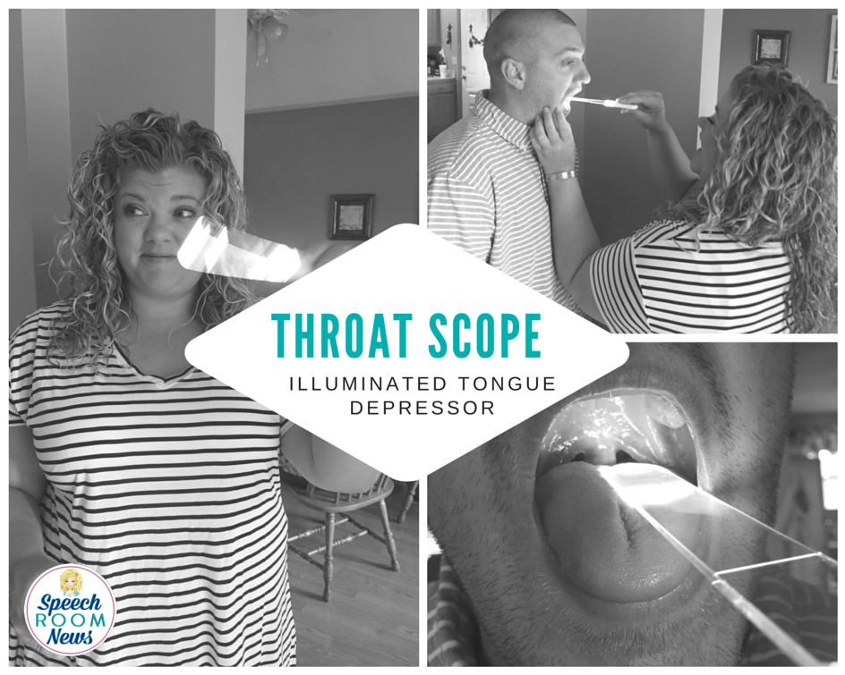
My brother was in town and I forced him to let me inspect his oral cavity using the throat scope. I was surprised at the amount of light the Throat Scope puts out. It definitely works!

The disposable blades make it perfect for healthcare.

If you want more details about the Throat Scope, visit their website.

This product was provided for review, no other compensation was provided and these opinion belong to me! And that’s my Throat Scope review :)

Read the original blog by Jenna at The Speech Room News HERE.

About the Author

My name is Jenna Rayburn Kirk, M.A.,CCC-SLP. I’m an ASHA certified pediatric speech-language pathologist. I work in the Columbus, Ohio area. I’m a graduate of The Ohio State University and Ohio University. My clinical experience has focused on pediatric speech and language therapy. I’ve worked with children aged 2-15 in schools and clinics. My current interest is in preschool-aged children. I work in a public school full-time in central Ohio.


Never miss a blog!

Subscribe to get updates on new blogs from Throat Scope.


Check Your Mouth: Oral Cancer Foundation’s Early Self Discovery Campaign

Check Your Mouth: The Early Self Discovery Campaign for Oral Cancer

The Oral Cancer Foundation (OCF) has launched a new nationwide campaign aimed at the early self discovery of oral cancers. The Check Your Mouth Campaign  is an interactive website designed to help individuals learn to self-discover suspicious tissue changes in their own mouths.

“Early discovery of oral cancers has profound implications. Individuals whose disease is found at early stages of development through opportunistic screenings have less invasive treatments, reducing treatment related morbidity, and there is also the opportunity for better long-term outcomes.”

-Dr. Ross Kerr, Oral Medicine Specialist at NYU

The new website, www.checkyourmouth.org, provides the knowledge and tools needed to recognize and find early symptoms of disease. The process is a very simple visual and tactile process. A person who finds suspect areas in their mouths that persists for over two weeks is then prompted to seek further evaluation from a dental professional.

With a step-by-step video, a very effective and inexpensive intra-oral light from Throat Scope, and examples of abnormalities to look for, this interactive campaign’s objective is to reduce late stage discovery of oral cancer. Throat Scope is available here.

About Oral Cancer

Oral cancer is incredibly deadly, killing one person in America every hour of every day, all year. In 2018, the foundation predicts there will be approximately 51,550 individuals diagnosed with an oral or oropharyngeal cancer.

Of those newly diagnosed today, only about 60% will be alive in 5 years.

The high fatality rate is largely due to late discovery of the disease.

To compound the problem, a national screening policy is not in place, and many individuals are not even aware that oral cancer is a significant and increasing common issue in the U.S.

“Reaching the general public with a message can be an expensive idea for a small nonprofit like OCF. Our partner organizations and the members they represent have been long-time leaders of performing oral cancer screenings as a part of the services they provide to patients. These organizations have been champions of routine visual and tactile screenings, and through ongoing advocacy efforts have made them part of the routine screenings dental hygienists perform.”

-Brian Hill, Executive Director of OCF, who is also a survivor of a late stage oral cancer

To help meet that challenge, the Oral Cancer Foundation has engaged two key strategic partners, the American Dental Hygienists’ Association (ADHA) and the Canadian Dental Hygienists Association (CDHA). The combined membership of these partners exceeds 200,000 dental hygienists in the U.S. and Canada.

Now the foundation looks to these long-term partners in screening to introduce their patient populations to the idea of self-exams at home between professional appointments. The foundations should also expose their patient population to the Check Your Mouth concept and website.

How Can Early Self Discovery Benefit You?

Self-examination has become a major part of the general cancer conversation. Other cancer organizations have created campaigns to encourage people to discover abnormalities early. Notable examples are a program to discover deadly melanoma skin cancers early, and self-exams through breast palpation for breast cancers. Check Your Mouth hopes to do the same with their early self discovery campaign.

With the measurable impact of early discovery in other forms of cancer, the Oral Cancer Foundation has moved forward with a similar objective. Oral cancers and their premalignant tissue changes are often easily visible with the naked eye. The model is not so different, with a learning curve that is short and simple to become effective, and an examination process that can be accomplished in about 5 minutes.

The OCF educates and encourages the public to learn the common risk factors mostly found in lifestyle choices such as smoking for oral cancer and avoid them. However, they cannot stop oral cancer from happening.

Clearly, finding the cancer early is the next best idea. Check Your Mouth makes early self discovery easy.

Check Your Mouth is a campaign with the objective of producing better long-term outcomes, which is a very tangible opportunity we can capitalize on today.

Read the original blog by the Oral Cancer Foundation HERE.